IRESite record type: plasmid_with_promoter_and_putative_IRES_without_translational_characterization
The shape of the nucleic acid molecule translated: linear
The quality of the mRNA/+RNA sequence: only_IRES_fragment
The mRNA/+RNA description:
In vitro T7 transcript used for structure probing experiments of ELG1 IRES. Transcription was done from PCR
template, prepared from pBiCELG1 vector using ELG1 forward primer with built-in T7 promoter sequence
(taatacgactcactatagggACTTTTGGTGGGCATTTA) and ELG1 reverse primer (cctcgagCTGGGCGGGGAATA).
The mRNA/+RNA sequence represented in the +DNA notation:
Warning: mRNA sequence when devoid of trailing 'A's is still not a substring of the plasmid sequence. Is it because an intron is spliced out? Stay calm then. :-)
Credibility of mRNA sequence: end-to-end_sequence_reverse_engineered_and_should_match_experiment
The name of the promoter used to express this mRNA: T7
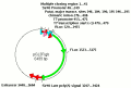
Description of the plasmid (facultative for promoter-less plasmid records): PCR template for structure probing experiments of ELG1 IRES was prepared from pBiCELG1 vector using ELG1
forward primer with built-in T7 promoter sequence (taatacgactcactatagggACTTTTGGTGGGCATTTA) and ELG1 reverse
primer (cctcgagCTGGGCGGGGAATA).
The in vivo produced transcripts are heterogeneous (due to any of promoter?/splicing?/cleavage?/breakage?): not tested
The in vivo produced heterogeneous transcripts occur due to alternative splicing: not tested
A promoter reported in cDNA corresponding to IRES sequence: not tested
The abbreviated name of the donor gene or virus from which this IRES was excised and inserted into the plasmid: ELG1
The IRES absolute position (the range includes START and STOP codons or their equivalents): 4-463
How IRES boundaries were determined: experimentally_determined
The sequence of IRES region aligned to its secondary structure (if available):
There is no Vienna RNA package installed on the server or some error/warning messages were output. Due to that maybe we cannot prepare 2D structures for display. The error/warning message was:
.......((.(..........(((..((..((..((((((((((((..(((....)))..))))))))))))..))..))..))))))...(((((((((((((((((.....(((((........))))).....))))))))))))))))).((((((.((((((((...(((((((((((((((((((((((.(((..((((..((..(....)..))..))))..))))))))))))))))))))))(((((.....)))))...........................................................)))).....))))))))))))))....................................((.....))......................(((..........)))..((.((......................
ERROR: unbalanced brackets in make_pair_table
STDOUT was:
Remarks:
ELG1 IRES represents part (from nt -803 to nt -461 of the original sequence) of 5' UTR from human ELG1 mRNA.
The absolute position of the experimentally mapped region (the range includes START and STOP codons or their equivalents): 4-463
The underlying nucleic acid sequence and structure of the mapped region:
There is no Vienna RNA package installed on the server or some error/warning messages were output. Due to that maybe we cannot prepare 2D structures for display. The error/warning message was:
.......((.(..........(((..((..((..((((((((((((..(((....)))..))))))))))))..))..))..))))))...(((((((((((((((((.....(((((........))))).....))))))))))))))))).((((((.((((((((...(((((((((((((((((((((((.(((..((((..((..(....)..))..))))..))))))))))))))))))))))(((((.....)))))...........................................................)))).....))))))))))))))....................................((.....))......................(((..........)))..((.((......................
ERROR: unbalanced brackets in make_pair_table
STDOUT was:
Remarks:
2D structure of ELG1 from Fig 5A.
3.1.1. Enzymes used to characterize at least partially the 2D structure.
Enzyme or a combination of enzymes used in a single experiment with respective buffer:
ss_experiment_with_enzyme_id: 58
The temperature (in degrees of Celsia): 22
The enzymatic method used to determine the 2D structure: ribonuclease T1
Enzyme or a combination of enzymes used in a single experiment with respective buffer:
Version: 0
pH 7.00
Li+ [mM] 0
Na+ [mM] 0
K+ [mM] 100.00
Mg2+ [mM] 10.00
Ca2+ [mM] 0
Cl- [mM] 120.00
Tris [mM] 10.00
BSA [mM] 0
HEPES [mM] 0
EGTA [mM] 0
EDTA [mM] 0
cacodylate [mM] 0
Enzyme or a combination of enzymes used in a single experiment with respective buffer:
ss_experiment_with_enzyme_id: 59
The temperature (in degrees of Celsia): 22
The enzymatic method used to determine the 2D structure: ribonuclease A
Enzyme or a combination of enzymes used in a single experiment with respective buffer:
Version: 0
pH 7.00
Li+ [mM] 0
Na+ [mM] 0
K+ [mM] 100.00
Mg2+ [mM] 10.00
Ca2+ [mM] 0
Cl- [mM] 120.00
Tris [mM] 10.00
BSA [mM] 0
HEPES [mM] 0
EGTA [mM] 0
EDTA [mM] 0
cacodylate [mM] 0
Enzyme or a combination of enzymes used in a single experiment with respective buffer:
ss_experiment_with_enzyme_id: 60
The temperature (in degrees of Celsia): 22
The enzymatic method used to determine the 2D structure: ribonuclease V1
Enzyme or a combination of enzymes used in a single experiment with respective buffer:
Version: 0
pH 7.00
Li+ [mM] 0
Na+ [mM] 0
K+ [mM] 100.00
Mg2+ [mM] 10.00
Ca2+ [mM] 0
Cl- [mM] 120.00
Tris [mM] 10.00
BSA [mM] 0
HEPES [mM] 0
EGTA [mM] 0
EDTA [mM] 0
cacodylate [mM] 0
Enzyme or a combination of enzymes used in a single experiment with respective buffer:
ss_experiment_with_enzyme_id: 61
The temperature (in degrees of Celsia): 22
The enzymatic method used to determine the 2D structure: ribonuclease T2
Enzyme or a combination of enzymes used in a single experiment with respective buffer: